
浏览量: 469
- 产品名称: Anti-GATA3 antibody
- 产品货号: CS1902-69
- 货期: 现货
- 价格与订购: 1500
- 数量:
库存: 100
- 规格: 50μL 100μL
- 产品信息
- 如何订购
产品描述
GATA3 is a transcription factor that in humans is encoded by the GATA3 gene. Studies in animal models and humans indicate that it controls the expression of a wide range of biologically and clinically important genes. The GATA3 transcription factor is critical for the embryonic development of various tissues as well as for inflammatory and humoral immune responses and the proper functioning of the endothelium of blood vessels. GATA3 haploinsufficiency (i.e. lose of one or the two inherited GATA3 genes) results in a congenital disorder termed the Barakat syndrome. Current clinical and laboratory research is focusing on determining the benefits of directly or indirectly blocking the action of GATA3 in inflammatory and allergic diseases such as asthma. It is also proposed to be a clinically important marker for various types of cancer, particularly those of the breast. However, the role, if any, of GATA3 in the development of these cancers is under study and remains unclear.
产品名称
Anti-GATA3 antibody
分子量
48 kDa
种属反应性
Human,Rat
验证应用
WB,IHC-P,FC
抗体类型
兔多抗
免疫原
Synthetic peptide within human GATA3 aa 1-100.
偶联
Non-conjugated
形态
Liquid
浓度
1 mg/mL
存放说明
Store at +4℃ after thawing. Aliquot store at -20℃. Avoid repeated freeze / thaw cycles.
存储缓冲液
IgG
纯化方式
Peptide affinity purified.
亚细胞定位
Nucleus.
数据链接
SwissProt: P23771 Human
其它名称
GATA 3 antibody GATA binding factor 3 antibody GATA binding protein 3 antibody
应用
WB:1:500-1:1,000 IHC-P:1:50-1:200 FC:1:50-1:100
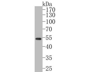
Fig1: Western blot analysis of GATA3 on SH-SY5Y cell lysates. Proteins were transferred to a PVDF membrane and blocked with 5% BSA in PBS for 1 hour at room temperature. The primary antibody (ER1902-69, 1/500) was used in 5% BSA at room temperature for 2 hours. Goat Anti-Rabbit IgG - HRP Secondary Antibody (HA1001) at 1:5,000 dilution was used for 1 hour at room temperature.
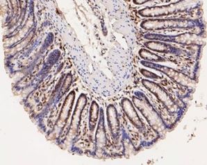
Fig2: Immunohistochemical analysis of paraffin-embedded rat large intestine tissue using anti-GATA3 antibody. The section was pre-treated using heat mediated antigen retrieval with Tris-EDTA buffer (pH 8.0-8.4) for 20 minutes.The tissues were blocked in 5% BSA for 30 minutes at room temperature, washed with ddH2O and PBS, and then probed with the primary antibody (ER1902-69, 1/50) for 30 minutes at room temperature. The detection was performed using an HRP conjugated compact polymer system. DAB was used as the chromogen. Tissues were counterstained with hematoxylin and mounted with DPX.
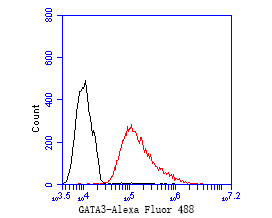
Fig3: Flow cytometric analysis of GATA3 was done on MCF-7 cells. The cells were fixed, permeabilized and stained with the primary antibody (ER1902-69, 1/50) (red). After incubation of the primary antibody at room temperature for an hour, the cells were stained with a Alexa Fluor 488-conjugated Goat anti-Rabbit IgG Secondary antibody at 1/1000 dilution for 30 minutes.Unlabelled sample was used as a control (cells without incubation with primary antibody; black).
背景文献
1. Perrino CM. et. al. Utility of GATA3 in the differential diagnosis of pheochromocytoma. Histopathology. 2017 Sep;71(3):475-479.
2. Asch-Kendrick R. et. al. The role of GATA3 in breast carcinomas: a review. Hum Pathol. 2016 Feb;48:37-47.
Note
For research use only .
 地 址:
地 址: 产品销售:
产品销售: E - mail :
 邮 编:
邮 编:
 Amily
Amily


